s-SNOM
provides nanoscale imaging and spectroscopy on all materials types across Vis, IR and THz
s-SNOM (scattering-type Scanning Near-field Optical Microscopy) delivers material-characteristic maps of chemical and optical properties of the sample surface at the spatial resolution of an atomic force microscope (AFM). Tuning the illumination wavelength, i.e. „color“ of the light source used for measurements provides spectroscopic information at the nanoscale.

Read the White Paper

s-SNOM is based on an asymmetric interferometer where the AFM tip and the sample are located in one of the interferometer arms. The light from the tip-sample arm is recombined with the reference beam at the detector. Patented interferometric detection based on varying the reference mirror position, thus reference phase, allows for simultaneous recording of phase and amplitude of the tip-scattered light, which relate to the local absorption and reflectivity, respectively. Patented modulaton of the reference phase and corresponding signal detection also enables complete suppression of the scattering background. In this way, s-SNOM returns pure optical/chemical near-field maps free of mechanical artifacts – all simultaneously with AFM topography and mechanical phase (size of the images above is 5x5 µm).
Technology Benefits
Highest versatility with proven performance on all material classes.
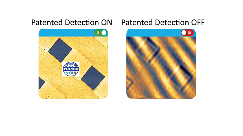
100% background suppression for reproducible artifact-free imaging and spectroscopy.
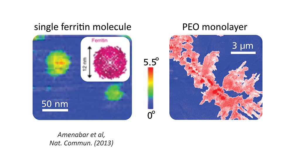
Best-in-class sensitivity capable of detecting single monolayers and individual macromolecules.
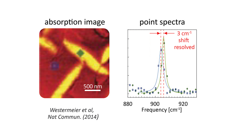
Nanoscale imaging & point spectroscopy for rapid component mapping and chemical identification.
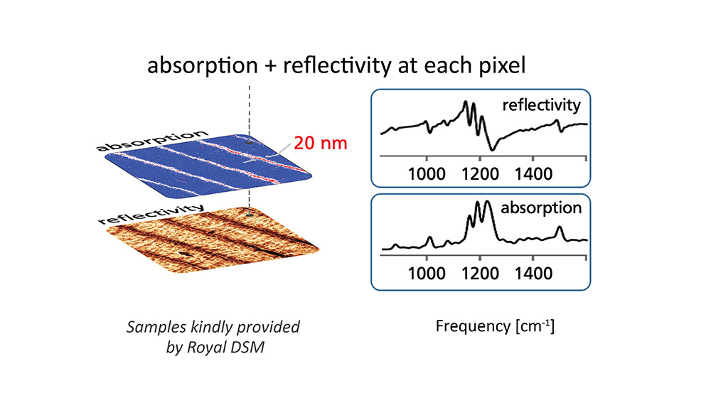
Simultaneous absorption & reflectivity at each pixel for complete characterization of sample’s optical properties with 10 nm spatial resolution.
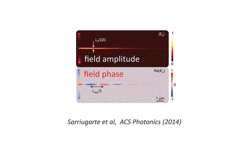
Electric field amplitude and phase mapping at 10 nm spatial resolution for complete modal analysis: attenuation, mode profile, wavelength, dispersion.
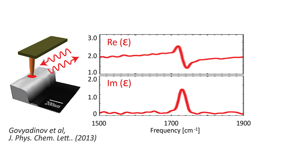
Access to the dielectric function at the nanoscale, i.e. refractive index and attenuation coefficient.
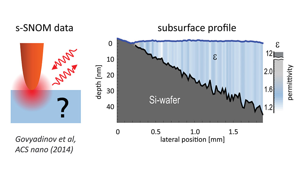
Quantitative subsurface analysis for complete chemometric analysis and characterization of sample structure.
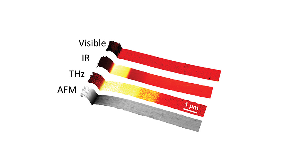
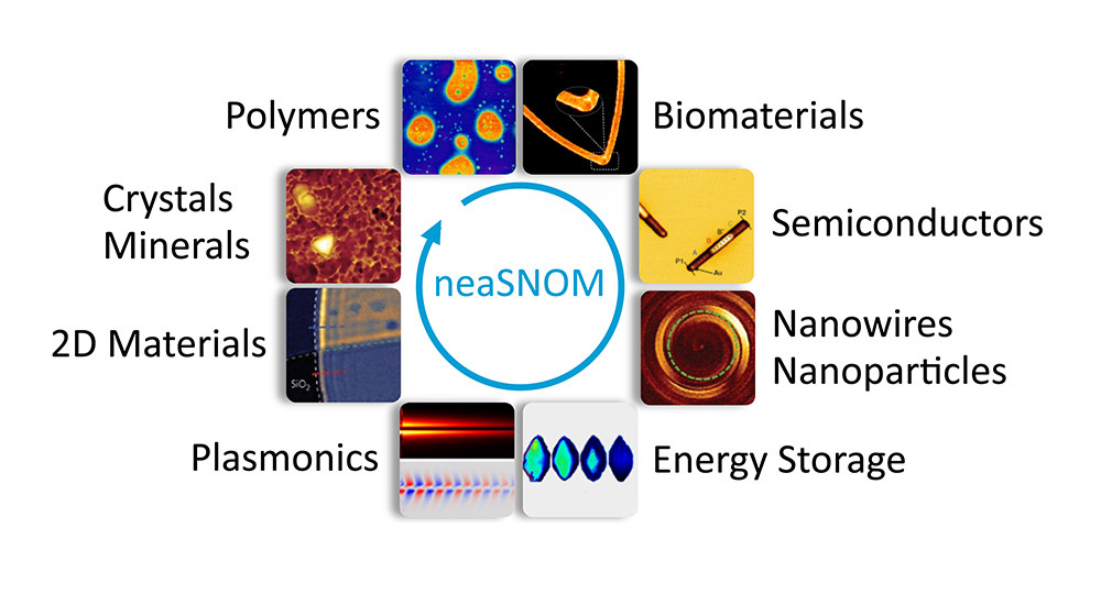
Nanoscale sample analysis at THz, IR and visible spectral ranges.
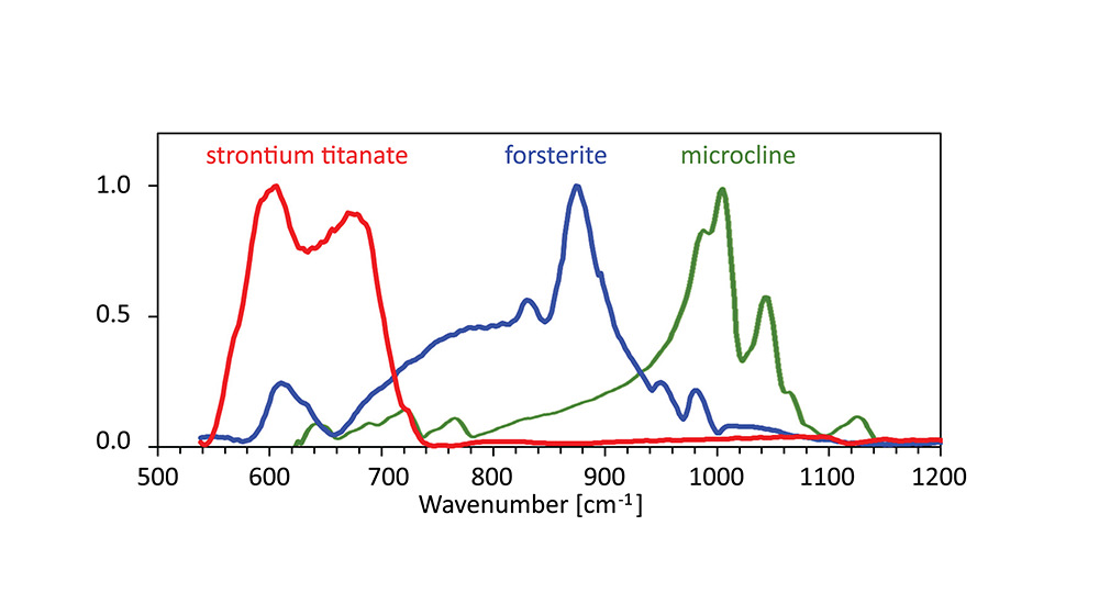
Exclusive s-SNOM point spectroscopy of low energy vibrations down to ca. 545 cm-1 for nanoscale analysis crystaline materials.